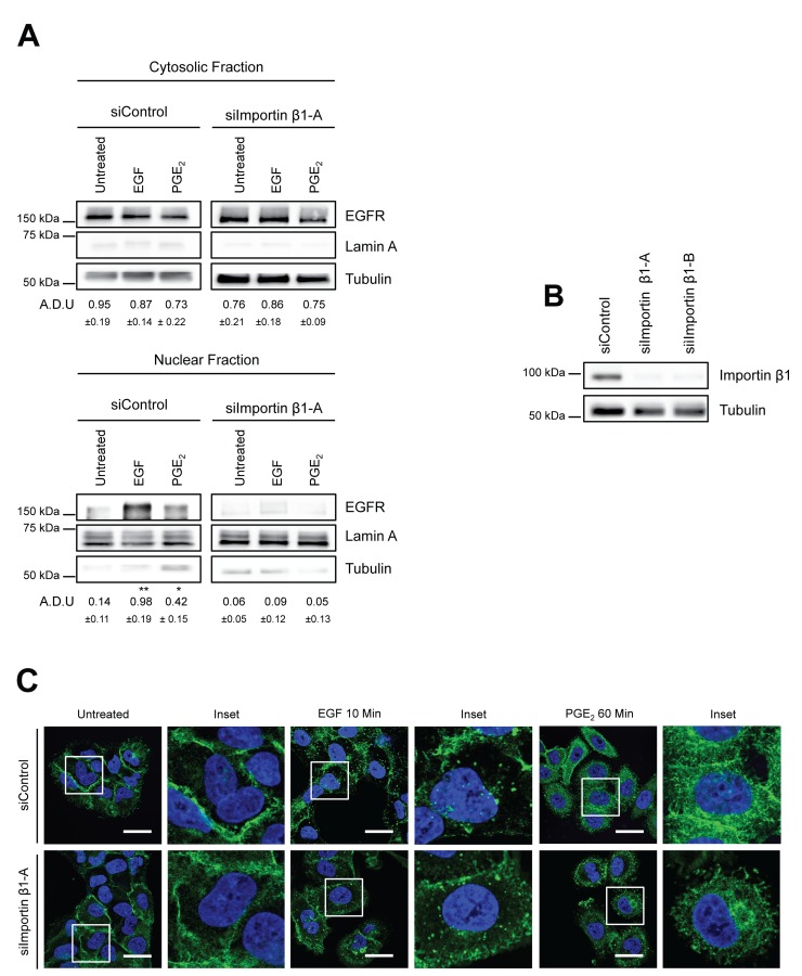
Figure 3. Importin β1 is required for EGFR nuclear translocation.
(A) A549 cells were transfected with siRNA control or siRNA against Importin β1 for 48h. Next, cells were serum starved overnight and exposed to 25ng/ml EGF for 10 min or to 1μM PGE2 for 60 min. EGFR level in cytoplasmic and nuclear fraction was assessed using immunoblot with indicated antibodies. (B) Knockdown efficiency was verified via western blot with Importin β1 antibody, Tubulin was used as loading control. Similar data were obtained with siImportin β1-B. Immunoblotting quantification was expressed in A.D.U. (arbitrary density unit) and as mean ± SD. *p < 0.05, **p < 0.01 vs Ctrl. EGFR in the cytoplasmic and nuclear fractions was normalized to Tubulin or Lamin A respectively. (C) A549 cells were transfected as indicated above. After that cells were fixed and stained for EGFR (green) and DAPI (blue). Confocal images were captured in the middle section of the nuclei with Leica SP5 confocal using 63x objective, scale bars 20 μm. Panel shows representative picture for each experimental condition. Boxed areas are shown in detail in the inset. The experiments were performed three times.

