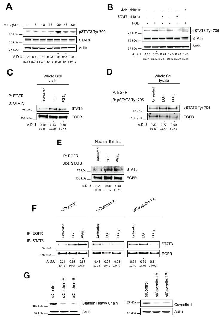
Figure 4. PGE2 induces the formation of EGFR-STAT3 complex into the nucleus.
(A) Immunoblotting analysis of STAT3 phosphorylation on Tyr 705 in overnight starved A549 exposed to 1 μM PGE2 for 5–60 min. (B) Immunoblotting analysis of STAT3 phosphorylation on Tyr 705 in A549 exposed for 30 min to 1 μM PGE2, with or without pre-incubation with JAK inhibitor, Ruxolitinib (10 μM), or STAT3 inhibitor, STAT3 inhibitor VII, for 30 min. Actin was used as loading control. (C–E) A549 cells were overnight starved and then exposed to 25ng/ml EGF or 1μM PGE2 for 10 and 60 min respectively. Whole cell lysate (C-D) and nuclear extract (E) were subjected to immunoprecipitation with anti-EGFR antibody and analyzed by immunoblotting with anti-STAT3 or anti-phosphoSTAT3 Try 705 antibodies. (F) A549 cells were transfected with siRNA Control or siRNA against Clathrin Heavy Chain or against Caveolin-1 for 24 h. Cells were then serum starved overnight and treated with 25ng/ml EGF for 10 min or 1μM PGE2 for 60 min. Whole cell lysates were subjected to immunoprecipitation with anti-EGFR antibody and analyzed by immunoblotting with anti-STAT3 antibody. (G) Knockdown efficiency was verified via western blot with Clathrin heavy chain and Caveolin-1 antibodies, actin was used as loading control. Data are shown only for siClathrin-A and siCaveolin-1A, similar data were obtained with siClathrin-B and siCaveolin-1B. Immunoblotting quantification was expressed in A.D.U. (arbitrary density unit) and as mean ± SD. *p < 0.05, **p < 0.01 vs Ctrl. In panel A and B, pSTAT3 Tyr 705 was normalized to STAT3. In panels C-F, STAT3 was normalized to EGFR. The experiments were performed three times.

