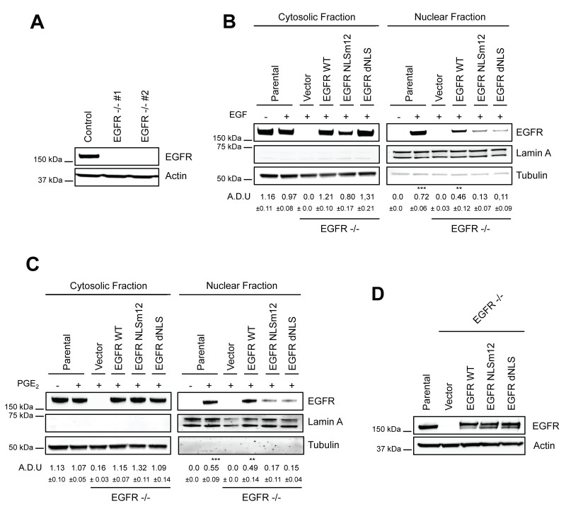
Figure 5. Generation and characterization of a model to study nuclear EGFR functions.
(A) Immunoblotting analysis of EGFR expression in A549 wild type cells and two clones knockout for EGFR, generated by CRISPR/Cas9 (EGFR -/- #1, #2). Actin was used as loading control. (B–C) EGFR knockout cells were transiently transfected with Vector or EGFR-WT or EGFR NLS mutant (NLSm12 or dNLS) plasmids for 48 h. Then EGFR nuclear import in response to 25 ng/ml EGF for 10 min (B) or to 1 μM PGE2 for 60 min (C) was analyzed by immunoblotting upon cell fractionation. Parental cells were included as a control. Tubulin and Lamin A were used as loading control for cytosolic and nuclear fraction respectively. Immunoblotting quantification was expressed in A.D.U. (arbitrary density unit) and as mean ± SD. *p < 0.05, **p < 0.01, ***p < 0.001 vs Ctrl. EGFR in the cytoplasmic and nuclear fractions was normalized to Tubulin or Lamin A respectively. (D) Expression of EGFR in EGFR -/- #1 cells, transfected with Vector, EGFR-WT and NLS mutant plasmids for 96 h. Actin was used as loading control. The experiments were performed three times.

