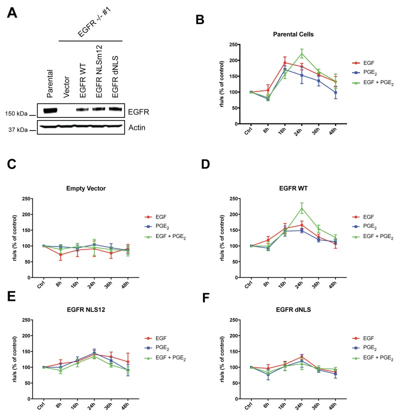
Figure 8. EGF and PGE2 induce nuclear EGFR-mediated A549 cell proliferation.
(A) Immunoblotting analysis of EGFR expression in A549 EGFR -/- #1 cells transfected with Vector, EGFR-WT and NLS mutant plasmids for 96 h. Parental cells were included as a control. Actin was used as loading control. (B–F) Parental cells or EGFR -/- #1, #2 cells transfected with Vector or EGFR WT or EGFR NLS12 or EGFR dNLS mutant plasmids for 24 h were harvested and seeded for BrdU incorporation assay. Cell proliferation was assessed by measuring the luminescence after 8, 16, 24, 36, 48 h treatment with EGF or PGE2 or the combination. Data are mean ± SD of triplicate cultures, expressed as % of control. Statistical analysis is reported in Table 3.

