. 2018 Mar 16;7:e31275. doi: 10.7554/eLife.31275

Figure 5. SPIN1 depletion increases ribosome-free uL18 and uL5.

(A) Knockdown of SPIN1 releases free forms of uL18 and uL5. HCT116p53+/+ were transfected with scramble or SPIN1 shRNA for 36 hr and subjected to sucrose gradient fractionation assay followed by WB analysis with indicated antibodies. (B) SPIN1 knockdown increases the endogenous uL18/uL5-MDM2 interaction. Cell lysates of HCT116p53+/+ cells transfected with scramble or SPIN1 shRNA were immunoprecipitated with MDM2 or control IgG, and analyzed by WB analysis with indicated antibodies. (C) SPIN1 overexpression counteracts p53 activation induced by ActD or 5-Fu. U2OS cells were transfected with pcDNA or Flag-SPIN1 for 48 hr, and treated with ActD or 5-Fu for 12 hr before harvested for WB analysis with indicated antibodies. (D) and (E) Knockdown of uL18 or uL5 compromises the induction of p53 by SPIN1 depletion. U2OS cells were transfected with scramble siRNA, SPIN1 siRNA, uL18 siRNA (D) or uL5 siRNA (E) as indicated for 48 hr. Cell lysates were subjected to WB analysis with indicated antibodies.

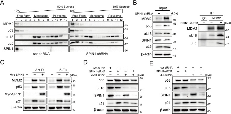
Figure 5.

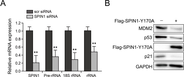
Figure 5—figure supplement 1. SPIN1 knockdown reduces rRNA expression and SPIN1-Y170A mutant retains activity to repress p53.

Figure 5—figure supplement 1.

(A) Scramble siRNA or SPIN1 siRNA was introduced into U2OS cells. RNA levels were analyzed using Q-PCR (*p value < 0.05; **p value < 0.01 by tailed t-test; n = 6). (B). SPIN1-Y170A inhibits p53. U2OS cells were transfected with control plasmids or FLAG-SPIN1-Y170A and the cells were collected for western blot analysis 48 hr after transfection.
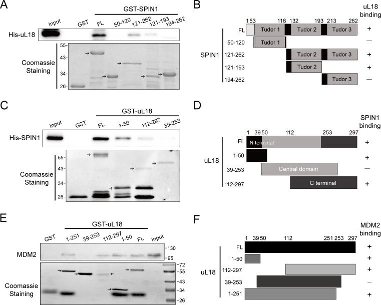
Figure 5—figure supplement 2. Mapping of domains responsible for uL18-SPIN1 and uL18-MDM2 binding.

Figure 5—figure supplement 2.

(A) uL18 interacts with the second Tudor like domain of SPIN1. Purified GST-tagged SPIN1 fragments, including aa 1-262(FL), aa 50–120, aa 121–262, aa 121–193, aa 194–262 and GST protein alone were incubated with purified His-uL18 protein for 3 hr at 4°C. Bound proteins were detected by WB analysis using anti-uL18 or coomassie staining. (B) A schematic diagram of uL18-binding regions on SPIN1 based on the result from (A). (C) SPIN1 interacts with both the N- and C-termini of uL18. Purified GST-tagged uL18 fragments, including aa1-297(FL), aa 1–50, aa 112–297, aa 39–253 or GST protein alone were rotated with purified His-SPIN1 protein for 1 hr at 4°C. Bound proteins were detected by WB analysis using anti-SPIN1 or coomassie staining. (D) A schematic diagram of SPIN1-binding regions on uL18 derived from the result from (C). (E) MDM2 interacts with both the N- and C-termini of uL18. Purified GST-tagged uL18 fragments, including aa1-297(FL), aa 1–50, aa 112–297, aa 39–253, aa 1–251 or GST protein alone were rotated with purified His-MDM2 protein for 4 hr at 4°C. Bound proteins were detected by WB analysis using anti-MDM2 (2A10) or coomassie staining. (F) A schematic diagram of uL18 binding regions on MDM2 based on the result from (E).