
Fig. 6. Cardiac fibroblast and MSC spread areas are dynamically tuned to PDMS stiffness.
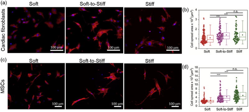
Representative images (a,c) and spread area quantification (b,d) of (top) cardiac fibroblasts and (bottom) MSCs on soft and stiff static substrates, as well as on a soft-to-stiff dynamic substrate on day 2 (stiffening performed on day 1). Stiff substrates were used as a positive control as photocrosslinking was applied before cell seeding and static soft substrates did not receive light exposure. The moduli for the Soft and Stiff substrates were ~3 and ~37 kPa, respectively. The modulus of Soft-to-Stiff substrate was changed from ~3 to 37 kPa after stiffening. Thus, the final moduli of the Soft, Soft-to-Stiff and Stiff substrates were ~3, ~37 and ~37 kPa, respectively. Cardiac fibroblasts were stained with vimentin (red), nuclei (blue); MSCs were stained with F-actin (red), nuclei (blue). n > 40 cells per group. ***: p < 0.001, n.s.: not significant. (For interpretation of the references to colour in this figure legend, the reader is referred to the web version of this article.)