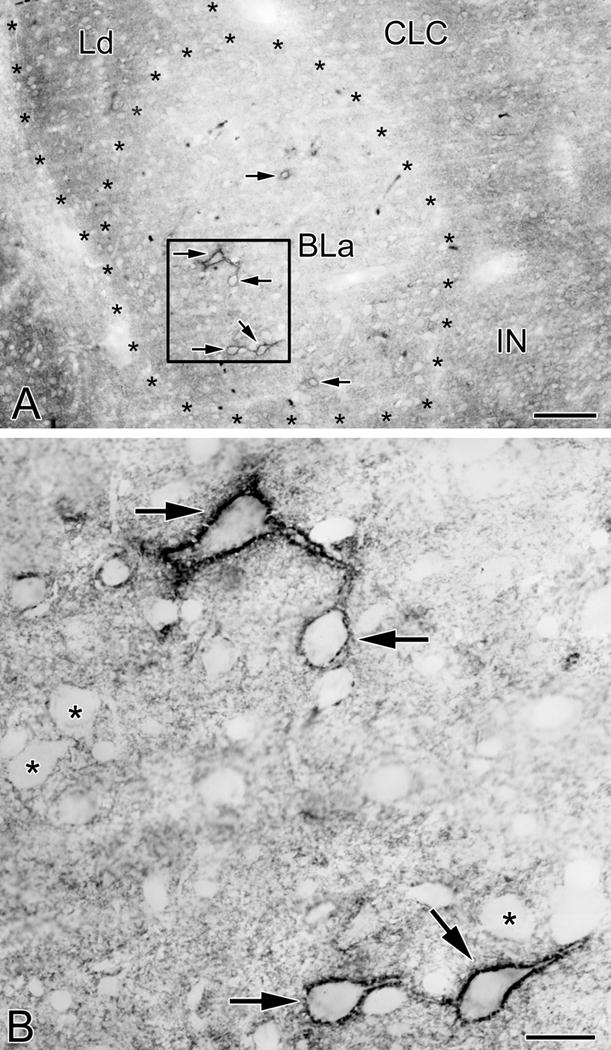
Fig. 1.

VC1.1-ir in PNNs and the neuropil in the anterior subdivision of the basolateral nucleus (BLa). (A) Photomicrograph of VC1.1-ir in the BLa at the bregma −1.8 level. Asterisks indicate the borders of BLa and the dorsolateral subdivision of the lateral nucleus (Ld). Four neurons ensheathed by VC1.1+ PNNs are in the boxed area of BLa (arrows; see B). Two additional BLa neurons ensheathed by VC1.1+ PNNs are also indicated by arrows. Other abbreviations: CLC, lateral capsular subdivision of the central nucleus; IN, intercalated nucleus. (B) Higher power photomicrograph of the boxed area in A. Note punctate VC1.1-ir in the neuropil and along the plasma membranes of the cell bodies and proximal dendrites of four neurons (arrows). In addition, some VC1.1-ir in the neuropil is in the form of small rings that are 1-2 μm in diameter. Asterisks indicate representative neurons that are not ensheathed by PNNs, but whose plasma membranes are decorated by punctate neuropilar VC1.1-ir. Scale bars = 100 μm in A, 20 μm in B
