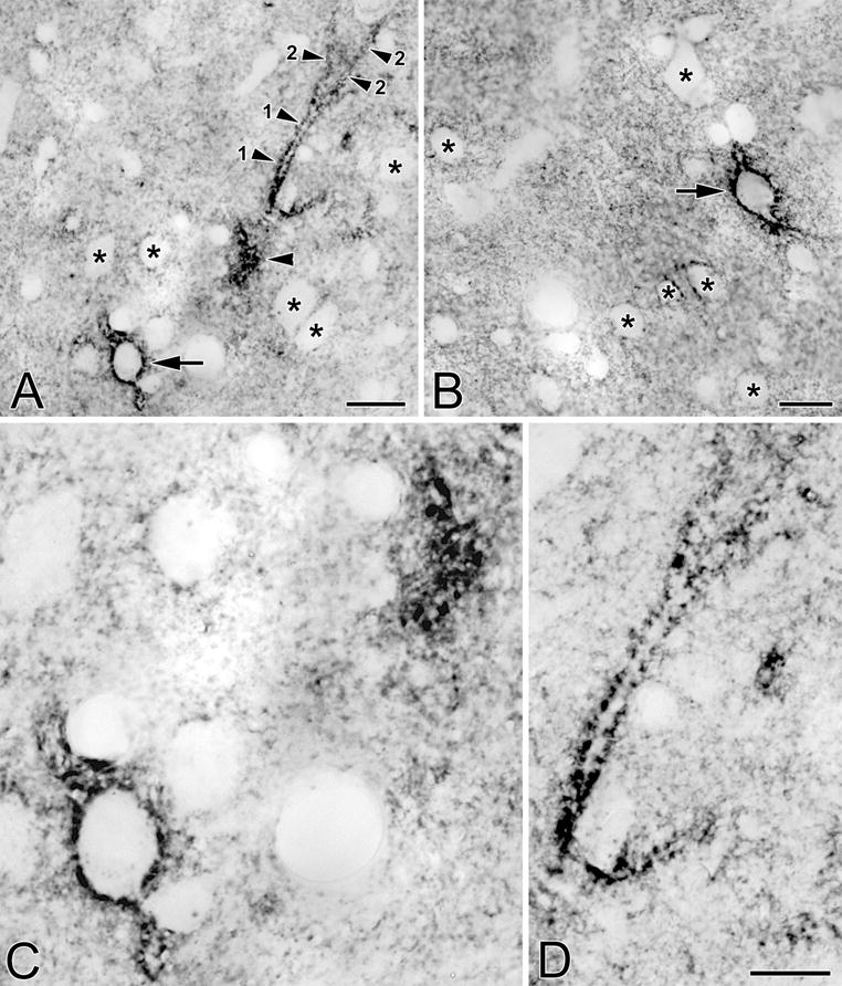
Fig. 3.

(A) Photomicrograph of a neuron in the BLa that is ensheathed by a VC1.1+ PNN (arrow). Also in the field is a primary dendrite (1) and two secondary dendrites (2) of another neuron. Only the edge of the cell body giving rise to these dendrites was in this section (unlabeled arrowhead). (B) Photomicrograph of a neuron in the lateral nucleus that is ensheathed by a VC1.1+ PNN (arrow). (C) Higher power view of the cell bodies in A. (D) Higher power view of the dendrites in A. Asterisks in A, B and C indicate representative neurons that are not ensheathed by PNNs, but whose plasma membranes are decorated by punctate neuropilar VC1.1-ir. Scale bars = 20 μm for A and B; 10 μm in D (C is at the same magnification)
