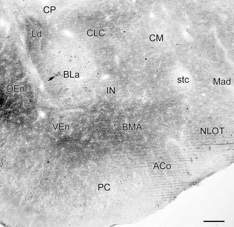
Fig. 5.

Photomicrograph of VC1.1-ir in a coronal section through the rostral amygdala (bregma −1.8 level; compare with Fig. 2A; see caption of Fig. 2 for abbreviations). Figure 1A is a higher power photomicrograph of the BLa in this section. Arrow indicates a large neuron in the anterior subdivision of the basolateral nucleus (BLa) that is ensheathed by a VC1.1+ PNN. There are 5 additional PNNs in the BLa in this section, and 3 in the nucleus of the lateral olfactory tract (NLOT), but they are difficult to see at this low magnification. Thus, almost all of the VC1.1-ir seen in this section is in the neuropil. Scale bar = 200 μm
