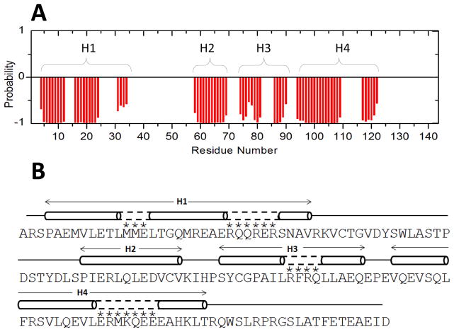
Fig. 2.
Primary and secondary structures of RD3. (A) TALOS+ ANN-secondary structure probability plotted as a function of residue number. (B) Secondary structure elements (cylinder for helix and line for random coil) were calculated on the basis of chemical shift index and sequential NOE patterns. Residues marked with an asterisk are not assigned but are predicted to have helical structure (dashed lines) based on a secondary structure prediction from the amino acid sequence.

