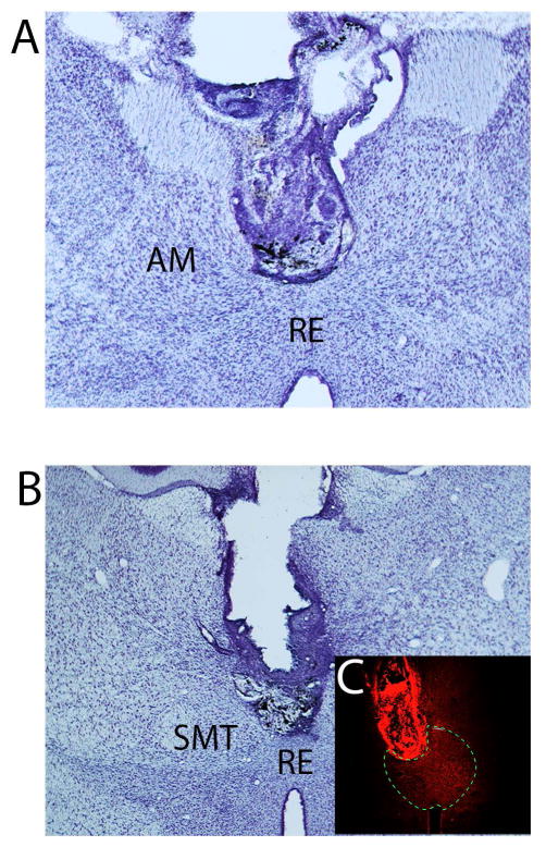
Figure 3.
Low magnification photomicrographs of Nissl stained transverse sections showing the locations of guide cannula in the rhomboid (A) and reuniens (B) nuclei of the ventral midline thalamus of two representative cases. C: photomicrograph illustrating the spread of the fluorescent conjugated muscimol restricted to RE and RH in this representative case. The borders of RE are indicated with the dashed line. Abbreviations: AM, anterior medial nucleus of the thalamus; RE, nucleus reuniens of thalamus; RH, rhomboid nucleus of thalamus; SMT submedial nucleus of thalamus.

