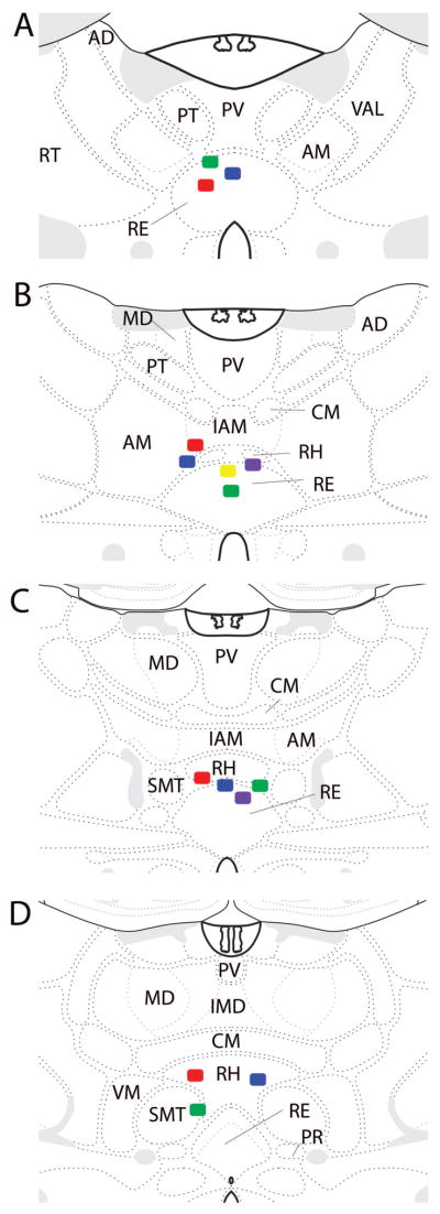
Figure 4.
Schematic diagrams illustrating the infusion sites at four levels (A–D) of the midline thalamus (plates adapted from Swanson, 2003). Colored squares show the position of the tip of the infusion cannula (1 mm below the end of guide cannula). As shown, infusion sites were located within the rhomboid (RH) and reuniens (RE) nuclei of the ventral midline thalamus in each of the included cases. Abbreviations: AD, anterior dorsal nucleus of thalamus; AM, anterior medial nucleus of thalamus; CM, central medial nucleus of thalamus; IAM, interoanteromedial nucleus of thalamus; IMD, interomediodorsal nucleus of thalamus; MD, mediodorsal nucleus of thalamus; PR, perireuniens nucleus of thalamus; PT, parataenial nucleus of thalamus; PV, paraventricular nucleus of thalamus; RT, reticular nucleus of thalamus; SMT, submedial nucleus of thalamus; VAL, ventral anterior lateral nucleus of thalamus; VM, ventromedial nucleus of thalamus.

