
Fig. 2.
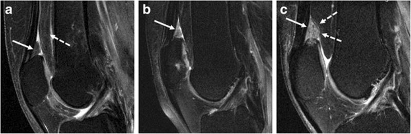
Representative sagittal fat-saturated intermediate-weighted fast spin-echo sequences (FOV, 160mm; slice thickness, 3mm; gap, 0mm; flip angle, 180°; TE/TR, 30/3200ms; acquisition time, 4.7mins) of the right knee in three subjects: A—Normal appearance of the SPFP with isointense signal of the SPFP (arrow) compared to the posterior suprapatellar (prefemoral) fat pad (dashed arrow) and without mass effect; B—hyperintense signal alteration of the SPFP (arrow); C—Hyperintense signal alteration (arrow) and mass effect with convex posterior border of the SPFP (dashed arrows).