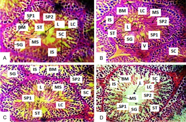
Figure 2.

Photomicrographs of a section of testis of Control (A), BS- (B), AA- (C) and BS+AA-treated rats (D). SG-Spermatogonium, L-Lumen, SP1-Primary spermatocyte, SP2-Secondary spermatocyte, BM-Basement membrane, MS-Mature spermatocyte, LC-Leydig cells, SC-Sertoli cells, ST-Spermatids and IS-Interstitium, V-Vacuole. (A) Showed normal testicular tissue; (B) Showed deleterious basement membrane, distorted spermatogenic cell and seminiferous tubule with lumen vacuolation; (C) Showed normal testicular tissue with hyperplasia of sertoli cells and (D) showed deleterious lumen, disrupted basement membrane, hyperplasia of sertoli cells, disruption of spermatogenic cells and degeneration of interstitium. (H&E paraffin stain; ×400, transverse section).
