Fig. 5.
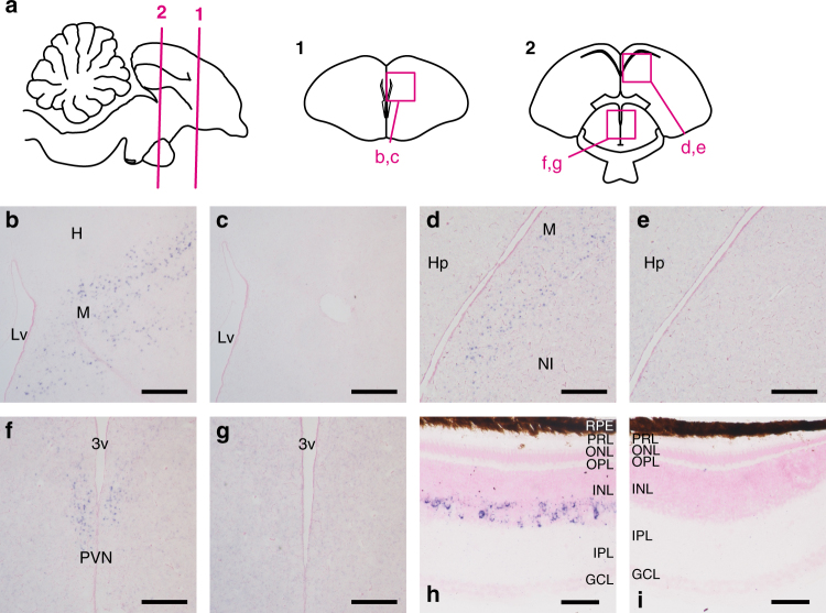
Distribution of Opn5L1 mRNA in chicken brain and retina. a Schematic drawings of chicken brain to show the approximate positions of frontal sections. Numbered lines in sagittal drawing indicate the positions of frontal sections numbered 1 and 2. Magenta boxes show the areas of panels b−g. b−g Opn5L1 mRNA in the chicken brain. Sections were hybridized with Opn5L1 antisense (b, d, f) and sense (c, e, g) probes. Panels (c), (e), and (g) show the tissue sections consecutive to (b), (d), and (f), respectively. h, i Opn5L1 mRNA in the chicken retina. Sections were hybridized with Opn5L1 antisense (h) and sense (i) probes. All the sections were counterstained with Nuclear Fast Red. Scale bars: b−g 300 μm; h, i 50 μm. Lv lateral ventricle, H hyperpallium, M mesopallium, Hp hippocampus, NI intermediate nidopallium, 3v third ventricle, PVN paraventricular nucleus, RPE retinal pigment epithelium, PRL photoreceptor layer, ONL outer nuclear layer, OPL outer plexiform layer, INL inner nuclear layer, IPL inner plexiform layer, GCL ganglion cell layer

