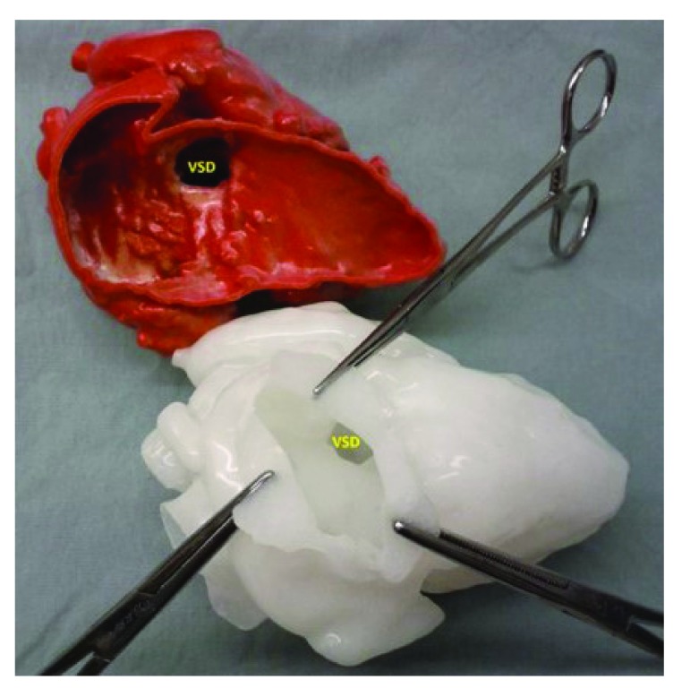
Figure 2. Three-dimensional modeling.
The upper image is a three-dimensional model obtained from a magnetic resonance angiogram with the free wall of the right ventricle cut away revealing the location of the ventricular septal defect (VSD); the lower model, made from a soft pliable material, shows the appearance of the VSD as seen through a virtual incision in the right atrium, as would be seen by the surgeon.

