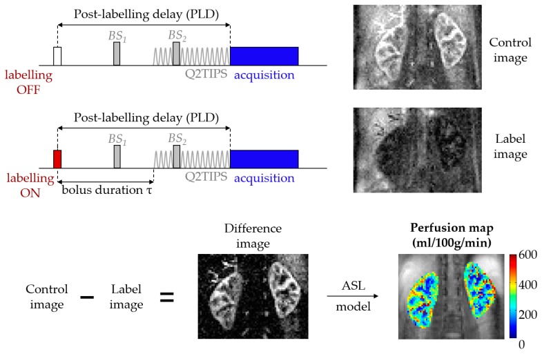
Figure 1.
Arterial spin labeling (ASL) overview. The background suppression (BS) pulses are optional but were used to acquire the renal ASL images in this example, hence the marked difference between control and labeled images (in healthy volunteers, the amount of signal due to inflowing blood is in the order of 5% of the non-background suppressed baseline tissue magnetization). The QUIPSS II with thin-slice TI1 periodic saturation (Q2TIPS) method [7] allows one to define the bolus duration in single post-labeling delay (single-PLD) pulsed ASL (PASL) studies.

