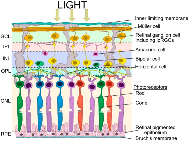
Figure 2.
Simplified schematic view of retinal layers involved in the pupillary light reflex. Vertical signalling pathways in the retina are composed of the photoreceptors (rod and cone cells), bipolar cells and retinal ganglion cells (RGC), including intrinsically photosensitive retinal ganglion cells (ipRGCs). There are also two lateral pathways comprised of horizontal cells in the outer plexiform layer (OPL) and the amacrine cells in the inner plexiform layer (IPL). These cells modulate the activity of other retinal cells in the vertical pathway. The somata of the neurons are in three cellular layers. The rod and cone cells are located in the outer nuclear layer (ONL), which is adjacent to the retinal pigment epithelium (RPE). The horizontal cell, bipolar cell and amacrine cell somas are located in the inner nuclear layer (INL), whilst the ganglion cell somata are located in the ganglion cell layer (GCL). The axon terminals of the bipolar cells stratify at different depths of the inner plexiform layer, which is subdivided into the OFF outer sublamina (where OFF bipolar cells terminate) and the ON inner sublamina (where ON bipolar cells terminate). There are also ON and OFF bands of melanopsin dendrites from the ipRGCs, but both lie outside of the ON and OFF cholinergic bands within the IPL. The bipolar cells are photoreceptor specific and the bipolar dendrites synapse exclusively with either rod or cone cells.

