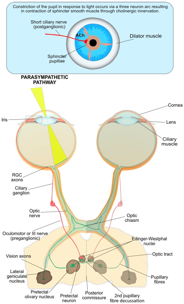
Figure 3.
The parasympathetic nervous system is the main system responsible for pupil constriction in response to light. The integrated afferent input is transmitted along the axons of the retinal ganglion cells (RGC), which contribute to the optic nerve. At the optic chiasm, nerves from the nasal retina cross to the contralateral side, whilst nerves from the temporal retina continue ipsilaterally. The pupillary RGC axons exit the optic tract and synapse at the pretectal olivary nucleus. Pretectal neurons are projected either ipsilaterally or contralaterally, across the posterior commissure, to the Edinger-Westphal nucleus. From there, the pre-ganglionic parasympathetic fibres travel with the oculomotor, or III cranial nerve, and synapse at the ciliary ganglion. The post-ganglionic parasympathetic neurons (short ciliary nerves) travel to and innervate the contraction of the iris sphincter muscle via the release of acetylcholine at the neuromuscular junction, resulting in pupil constriction.

