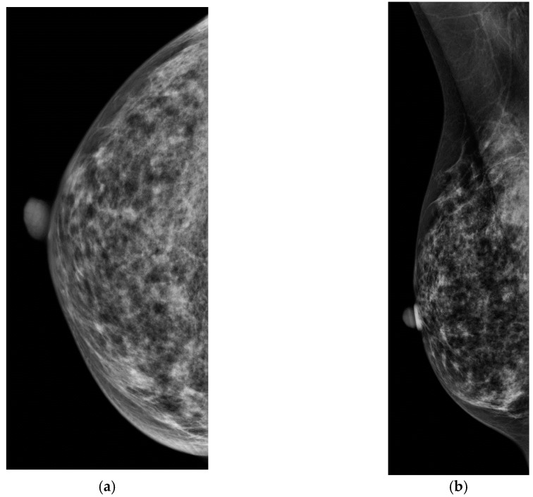
Figure 4.
(a,b) CC and MLO digital mammograms in a 56 year old woman with heterogenously dense breasts with an occult breast cancer. Automated whole breast ultrasound (ABUS). (c) transverse image of ABUS and (d) reconstructed coronal image demonstrate an irregular hypoehoic mass. (e) Handheld ultrasound confirms a 0.8 cm irregular, heterogenous hypoechoic mass. Pathology demonstrated mixed invasive ductal and invasive lobular carcinoma, grade 2, ER positive, PR and HER2 negative.


