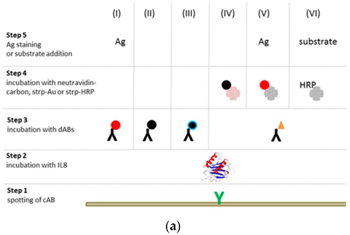
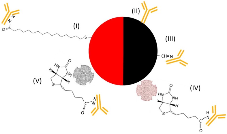
Figure 1.
(a) Schematic presentation of six distinct assay formats for colorimetric detection: (I) cAB + IL8 + gold-labeled dAB, (II) cAB + IL8 + carbon-labeled dAB, (III) cAB + IL8 + oxidized carbon-labeled dAB, (IV) cAB + IL8 + biot. dAB + neutravidin–carbon, (V) cAB + IL8 + biot. dAB + strp–gold, and (VI) cAB + IL8 + biot. dAB + strp-HRP + enzyme substrate. Slides treated via gold nanoparticles were further stained by silver (Ag). (b) Illustration of the antibody (in orange) coupling process using gold (red) and carbon (black) nanoparticles.


