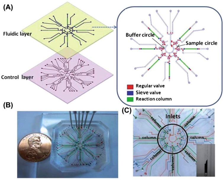
Figure 4.
(A) Schematic representation of fluidic layers of the immunoreaction chip used in the detection of algal toxins. Valves and columns are clarified by different colors: red (grey in print versions) for regular valves (for isolation), blue (dark grey in print versions) for sieve valves (for trapping protein A beads loaded in the column module) and green (light grey in print versions) indicates the immune columns by loading of microspheres. (B) Optical micrograph of the microfluidic chip. The various channels have been loaded with food dyes to help visualize the different components of the microfluidic chip: control line colors are as in (A), plus green (light grey in print versions) for fluidic channels. A penny coin (diameter 18.9 mm) is shown for size comparison. (C) Optical micrograph of the central area of the chip containing seven immunoreaction columns. Inset: a snapshot of the protein A beads loading process in action [22].

