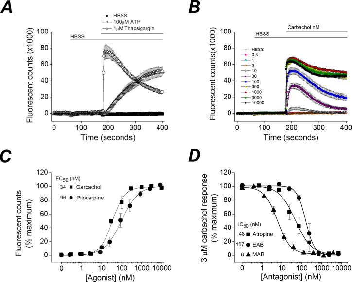
Figure 2.

Ethylatropine bromide inhibits the human M1 receptor. (A) Ca2+-dependent fluorescent signal of CHO-hM1 cells loaded with Fluo-4 was monitored. With use of the integrated fluidics of the FLEXStation II, a bolus of HBSS was delivered at 60 s, resulting in a miniscule reduction in the fluorescent signal. Addition of ATP (100 μM) or thapsigargin (1 μM) at 180 s resulted in a long-lasting increase in [Ca2+]i. Data are the mean of eight replicates from a single plate. Bars indicate the duration of drug exposures. (B) A rapid increase in fluorescence was detected after application of the muscarinic receptor agonist carbachol at various concentrations. Data are the mean of eight replicates from a single plate for each concentration applied. (C) Concentration–response curves of the carbachol and pilocarpine-induced Ca2+ signal. The maximum Ca2+ signal was measured and the response was calculated for increasing concentrations of each agonist (carbachol, EC50 = 34 nM, n = 3 experiments in replicates of 8; pilocarpine, EC50 = 96 nM, n = 5 experiments in replicates of 8). (D) Carbachol induces a rapid rise in intracellular Ca2+ at 3 μM, which can be attenuated in a concentration-dependent manner by pretreatment with atropine (IC50 = 48 nM, n = 3 experiments in replicates of 4), methylatropine bromide (MAB) (IC50 = 6 nM, n = 7 experiments in replicates of 4) or ethylatropine bromide (EAB) (IC50 = 157 nM, n = 3 experiments in replicates of 4), but not by HBSS (not shown). In (C) and (D), data are mean ± SEM.
