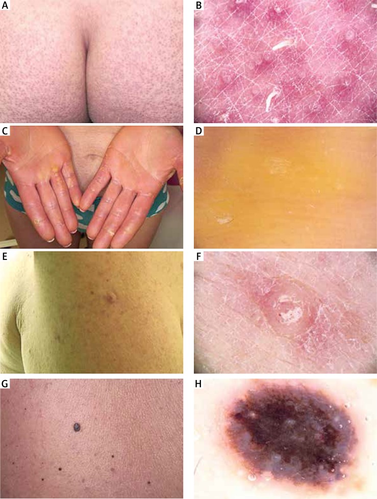
Figure 2.
Clinical and dermoscopic images of patients developing skin toxicities during vemurafenib therapy. Clinical picture of exacerbated keratosis pilaris in patient 7 (A). Dermoscopy revealed multiple hyperkeratotic intrafollicular filiform plugs within normal visible pilosebaceous orifice (B). Clinical picture of palmar‑plantar erythrodysaesthesia (G2) in patient 8 (C). Dermoscopy revealed hyperkeratotic skin changes in the form of yellowish, confluent, homogeneous masses in patient 8 (D). Clinical picture of proliferative, hyperkeratotic verruca in patient 5 (E). Dermoscopy showed several dotted vessels in an exophytic, amorphous proliferation (F). Clinical picture of suspected melanocytic lesion appeared as an “ugly duckling” lesion in patient 6 (G). Dermoscopically it was a lesion with a confluent, bluish-blackish homogeneous pattern suggesting melanoma, histopathologically identified as a blue naevus in patient 6 (H)

