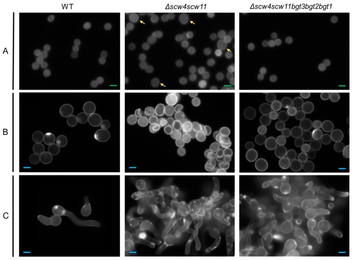
Figure 4.
Morphology of the dormant (A) 0 h; swollen (B) 4 h; and germinated (C) 8 h conidia of the parental strain, and Δscw4Δscw11 and Δscw4scw11bgt3bgt2bgt1 mutants using fluorescence microscopy (×63) with UV light and Calcofluor white® staining, in 3% Glucose medium +1% yeast extract. Green scale bars (2 µm in panel A), blue scale bars (6 µm in B and C), and yellow arrows highlight dormant conidia with abnormal size.

