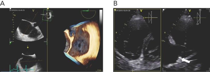
Figure 3: Pre-procedural Assessment of the Tricuspid Valve by Echocardiography.
A: Transoesophageal 3D echocardiography providing a ‘surgical view’ of the tricuspid valve in a patient with functional TR; the image illustrates a substantial central coaptation defect of the tricuspid valve leaflets due to dilatation of the tricuspid annulus. B: Imaging of the posterior portion of the tricuspid annulus by transthoracic X-plane echocardiography; the tricuspid valve is visualised in a modified parasternal short-axis view and the depth of the landing zone (arrow) for the Trialign device is measured on biplane imaging.

