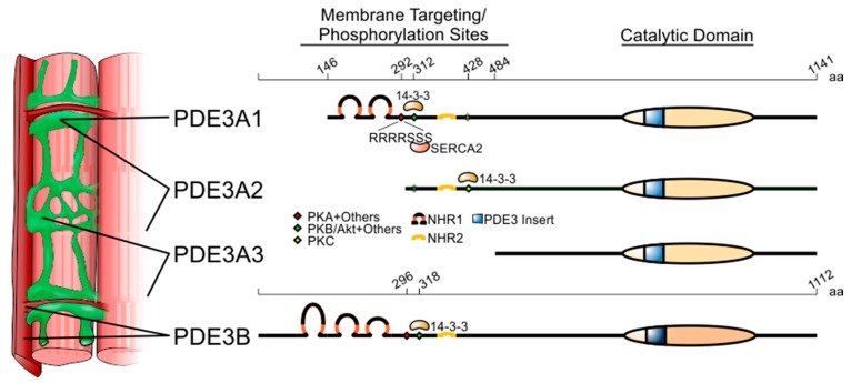
Figure 1.
Structure and subcellular localisation of the PDE3 genes and their variants. Length in amino acids (aa) is provided at the top of the two PDE3 isoforms. PDE3A1 is translated from the second AUG codon of the open-reading frame found in the PDE3 mRNA. While the longest variant of PDE3A, PDE3A1, is mainly localised to the sarcoplasmic reticulum, PDE3A2 and PDE3A3 are found both in membranes and cytoplasm. PDE3B is mainly localised to plasma membrane invaginations known as T tubules. Coloured diamonds indicate phosphorylation sites. Selected PDE3-interacting proteins are listed where the precise binding sequences are known. Membrane-associated N-terminal hydrophobic regions 1 and 2 (NHR1 and 2) are depicted as loops. The catalytic domain, highly conserved between PDE3A and PDE3B, is indicated as a striped oval that includes the 44-amino-acid insert characteristic of PDE3 isoforms.

