Fig. 2.

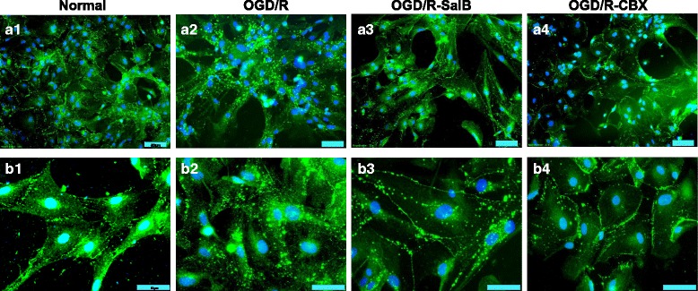
Redistribution of astrocytic Cx43 after OGD/R injury and the effects of SalB and CBX. We cultured primary astrocytes and performed cytoimmunofluorescent staining for Cx43. a1 In the normal group, Cx43 was mainly expressed discontinuously in the plasma membrane. b1 At high magnification, Cx43 mainly expressed at the gap junction and some were punctate distributed. a2, b2 In the OGD/R group, Cx43 was mainly expressed in the cytoplasm, which existed in the shape of block and grain. a3, b3 Compared to the OGD/R group, the OGD/R-SalB group exhibited weaker cytoplasmic Cx43 staining but enhanced plasma membrane Cx43 staining. The Cx43 expressed at gap junctions was morphologically similar to that in the normal group. a4, b4 The OGD/R-CBX group exhibited staining results similar to those of the OGD/R-SalB group, though the Cx43 at gap junctions covered a larger area than in the control and OGD/R-SalB groups. Scale bar = 50 μm
