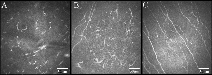
Figure 2.
In vivo confocal microscopy images of the subbasal corneal nerve plexus in eyes with herpes zoster ophthalmicus (HZO) and controls. Both the eyes affected by HZO (A) and the contralateral clinically unaffected eyes (B) demonstrated a significant reduction in subbasal nerve plexus including number of nerves, branches, and total nerve length when compared to normal controls (C). Reprinted from [51], with permission from Elsevier®.

