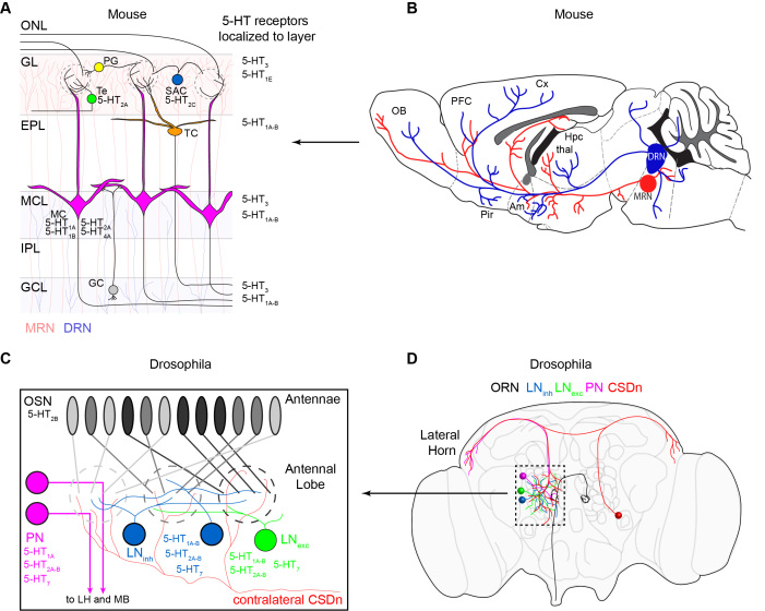
Figure 1.
The organization of the olfactory and serotonergic systems in mice and flies. (A). A schematic of the layers and major cell types in the mouse olfactory bulb (OB). Olfactory input comes from OSN axons in the olfactory nerve layer (ONL). OSNs synapse onto mitral cell (MC, pink) dendrites in glomeruli in the glomerular layer (GL). The mitral cells project out of the OB and to third order olfactory regions such as the piriform cortex (pir) and the amygdala (am). Juxtaglomerular neurons such as short axon cells (SAC, blue), periglomerular cells (PG, yellow), and external tufted cells (Te, green) modify olfactory processing in the glomerular layer. SACs and PG are inhibitory and Te cells are excitatory. Granule cells (GC, gray) in the granule cell layer (GCL) send dendrites to the external plexiform layer (EPL) where they make dendrodendritic synapses with MC. Serotonin receptors that have been attributed to each cell type are listed next to that cell. Serotonin receptor classes that have only been localized to layers of the bulb are listed next to that layer on the right. The median raphe nucleus (MRN) provides serotonergic innervation for the GL (light red shaded layer), and the dorsal raphe nucleus (DRN) provides innervation for the mitral cell layer (MCL) and granule cell layer (GCL) (light shaded blue). The most densely innervated layer is the GL. (B). The DRN projects from the brainstem to three major olfactory areas, the OB, the piriform (pir), and the amygdala (Am). MRN projections to the OB also arise from the brainstem. PFC = prefrontal cortex, Cx = cortex, HPC = hippocampus, thal = thalamus. Adapted from Muzerelle et al. 2016 with permission. (C). A schematic of the Drosophila antennal lobe (AL). OSNs residing on the antennae each project to a single glomerulus in the AL. Each OSN expressing the same receptor (denoted as the same shade of gray) project to the same glomerulus where they make synapses onto projection neurons (PN, pink). PNs send axons out of the AL to the lateral horn (LH) and mushroom body (MB) where third order olfactory processing takes place. Inhibitory (blue) and excitatory (green) local interneurons also influence glomerular processing in flies. Only two cells (one from each hemisphere) provide serotonergic innervation for the AL. These are the CSDns. (D). A schematic of the Drosophila brain showing the location and projections of the major cell classes discussed in this review.

