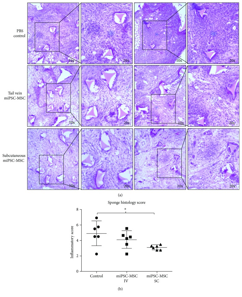
Figure 4.
In vivo assessment of the immunomodulatory properties of miPSC-MSC in a sponge model of periodontitis. (a) Representative light microscopy images of H&E stained sections of the implanted sponges 14 days after the injection of PBS, or 5 × 10⁶ miPSC-MSC via tail vein injection (IV) or subcutaneous injection (SC). (b) Semiquantitative scoring of the level of immune cell infiltrate present in histological slides of the implanted sponges (∗p value <0.05 as determined by one-way ANOVA with multiple comparisons, n = 6).

