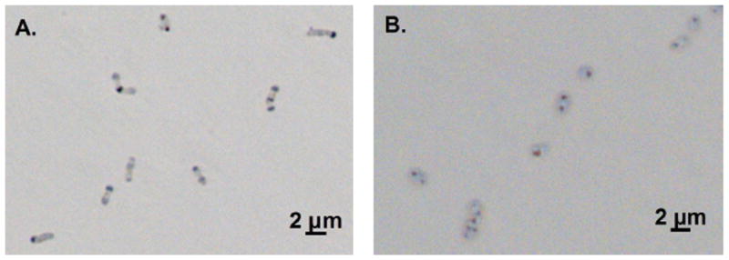
Figure 3. Polyphosphate storage by LM-2 and LM-P.

Polyphosphate granules were synthesized during and after P-starvation by LM-2 (A) and LM-P (B). Photos were taken 21.5 and 24.75 hours after transfer into medium with no additional P, respectively. Polyphosphate was stained with methylene blue and appears as dark spots in cells visualized with brightfield microscopy.
