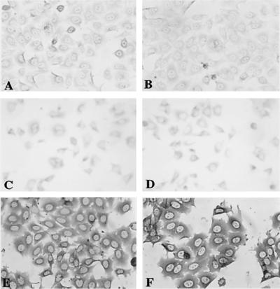
Figure 3.
Immunoperoxidase staining for α-amylase and lactoferrin. HSYzeo and HSYR2/R1 exhibited weakly positive staining for α-amylase (A and C) and lactoferrin (B and D). HSYR2-IIIb showed intensive positive staining for α-amylase (E) and lactoferrin (F) in the cytoplasm and perinuclear regions. The cells shown are representative of seven clones each.

