Figure 5.
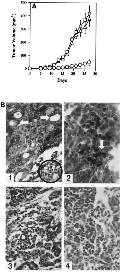
Analysis of tumors formed in nude mice. (A) The tumorigenicity of HSYzeo, HSYR2/R1, and HSYR2-IIIb. There was no significant difference between the tumor growth of HSYzeo (□) and HSYR2/R1 (▵) (P > 0.05). In contrast, there was a significant reduction in growth rate of HSYR2-IIIb-derived tumors (○) as compared with those of HSYzeo- and HSYR2/R1-derived tumors (P < 0.001). For each group, the mean ± SE was plotted. (B1) Tumors developed from HSYR2-IIIb exhibited predominant gland-like and acinar-like structures surrounded by stromal cells (arrowhead). In some areas, apoptotic bodies were observed in the nuclei of the tumor cells (circled) (original magnification: ×100). (B2) On TUNEL examination, tissue section of HSYR2-IIIb-derived tumor showed positive signals in tumor cells (arrowhead) (original magnification: ×200). The histological appearances of HSYR2/R1-derived (B3) and HSYzeo-derived (B4) tumors exhibited typical adenocarcinomas with a solid and trabecular pattern (original magnification: ×100).

