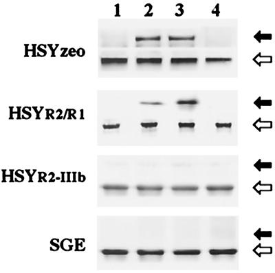
Figure 6.
FGF-stimulated phosphorylation of FRS2. The cell lysates extracted from SGE, HSYzeo, HSYR2/R1, and HSYR2-IIIb were immunoprecipitated with anti-Grb2 antibody and were blotted with anti-phosphotyrosine antibody or anti-FRS2 antibody. Closed arrowheads indicate the position of the phosphorylated form of FRS2, and open arrowheads indicate the position of the unphosphorylated forms of FRS2. Lane 1: Control; lane 2: FGF-1 stimulated; lane 3: FGF-2 stimulated; lane 4: KGF stimulated. Data represent the results of clone 7A and clone 7B, as other clones gave essentially the same results.

