Figure 6.
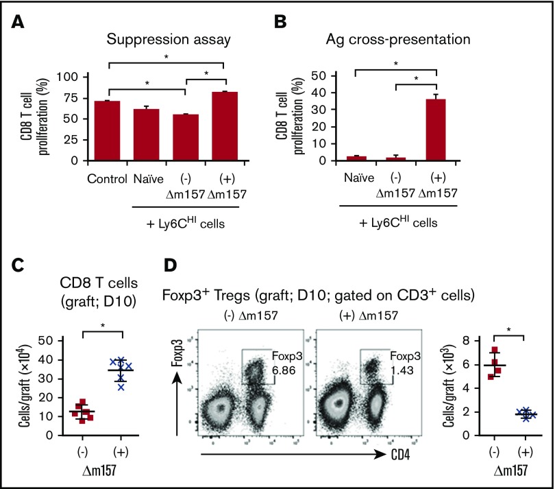
Functional assessment of Ly6CHIcells and intragraft CD8 T cells and CD4+Foxp3+Tregs. (A) In vitro suppression assay using sorted splenic Ly6CHI cells from the indicated groups 10 days posttransplantation. Control: no Ly6CHI cells were added. Sorted splenic Ly6CHI cells were cocultured with CFSE-labeled syngeneic CD8 T cells stimulated with anti-CD3/CD28 coated beads (at a ratio of 1:1:1). Proliferation of CD8 T cells was quantified by CFSE dilution. Data shown were averaged from a total of 4 mice in each group from 2 independent experiments. (B) Alloantigen cross-presentation by Ly6CHI cells to CD8 T cells. Sorted splenic Ly6CHI cells from the indicated groups were cocultured with CFSE-labeled naïve B6 CD8 T cells at a ratio of 5:1 (Ly6CHI:CD8) in the presence of BALB/c splenocyte lysates (50 µg/mL). CD8 T-cell proliferation was measured by CFSE dilution on day 4 of cocultures. Data shown in panels A and B were averaged from a total of 4 mice in each group from 2 independent experiments and presented as mean ± SD. *P < .05. (C) Quantitative analysis of graft-infiltrating CD3+CD8+ T cells (day 10 posttransplant) from the indicated groups. (D) Representative FACS plots demonstrating graft-infiltrating CD4+Foxp3+ Tregs (day 10 posttransplant; gated on CD3+ cells) from the indicated groups. Scatter graph showing quantitation of Treg numbers. Data shown in panels C and D were from 2 to 3 independent experiments with a total of 4 to 6 grafts in each group. Data were presented as mean ± SD. *P < .05.

