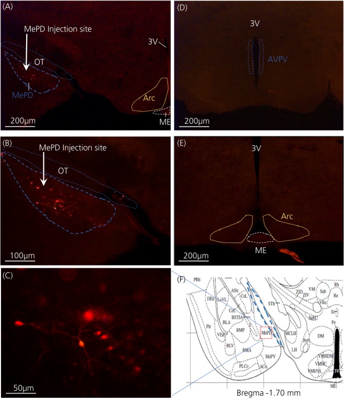
Figure 1.

Expression of posterodorsal medial amygdala (MePD) kisspeptin neurones with hM3Dq‐mCherry in Kiss‐Cre mice. Coronal section shows red mCherry fluorescence positive neurones (blue line) in the MePD but not in the arcuate nucleus (ARC) (A) and the white arrow indicates the injection site of AAV‐hSyn‐DIO‐hM 3D(Gq)‐mCherry into the MePD of Kiss‐Cre mice in the same section (B). Higher‐power view shows the MePD kisspeptin neurones tagged with mCherry (red fluorescence), which indicates hM3Dq receptor expressing kisspeptin neurones (C). The absence of red fluorescence in the anteroventral periventricular nucleus (AVPV) (blue dotted line) (D) and arcuate nucleus (ARC) (yellow dotted line) (E) shows the specificity of the viral contruct to MePD kisspeptin neurones. Schematic representation27 of MePD and its spatial relationship with the optic tract (blue dotted line) (F). ME, median eminence; OT, optic tract; 3V, third ventricle
