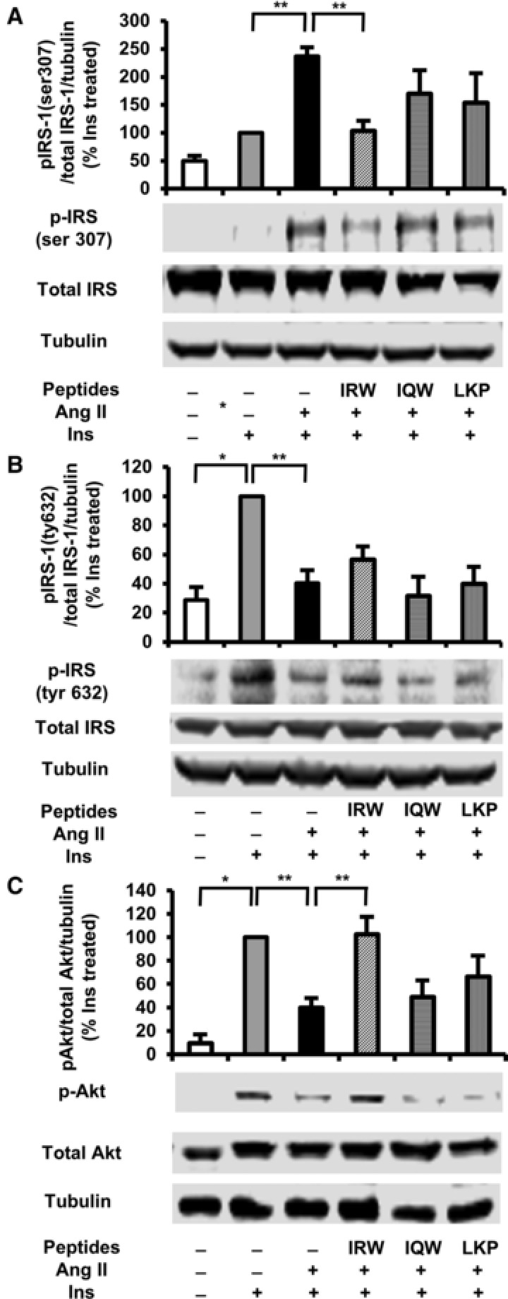
Figure 2.

Effects of egg white ovotransferrin‐derived tripeptides on insulin signaling pathway in Ang II treated L6 myotubes. Fully differentiated myotubes were treated with 100 μm of peptides for 2 h followed by treatment with 1 μm of Ang 2 for 24 h. L6 myotubes were preincubated in KHH buffer for 2 h. They were then incubated in KHH buffer containing 11 mm glucose without or with 100 nm of insulin for 30 min. The cells were lysed and western blotting of the lysates was performed with antibodies against p‐IRS‐1 Ser 307 (A), p‐IRS‐1 tyr 632 (B), total IRS‐1 (A,B), p‐Akt (C), total Akt (C), and α‐tubulin (loading control). A set of representative images is shown. Data are presented as mean ± SEM of three independent experiments. **indicates p < 0.05 as compared to the Ang II‐treated group. *indicates p < 0.05 as compared to the untreated group.
