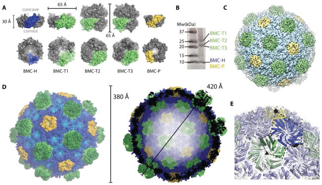
Fig. 1.
Overview of the components and the overall structure of the BMC shell. (A) Surface representation and dimensions of a side view (top row) and of the concave face (bottom row) of the structures of hexameric BMC-H (blue), trimeric BMC-T (lime) and pentameric BMC-P proteins (yellow) that constitute the shell. The BMC-T2 and BMC-T3 proteins each consist of two closely appressed pseudohexamers. The BMC-P structure was extracted from the whole shell structure, BMC-H and BMC-T1 from previously determined crystal structures (PDB IDs 5DJB and 5DIH, respectively) and BMC-T2 and BMC-T3 are crystal structures determined in this study. (B) SDS-PAGE of purified H. ochraceum BMC shells. (C) Overview of the 8.7 Å cryo electron microscopy structure colored by shell protein. (D) Surface representation of the crystal structure with a color gradient by distance from center (light to dark from inside to outside) (left) and cross-section through the center (right). (E) Close-up of the icosahedral asymmetric unit (dashed line) with symmetry axes indicated with solid symbols, pseudo threefold symmetry with open triangles. Only one stack shown for the BMC-T.

