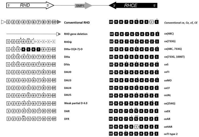
Figure 3.
Inverted orientation of the RHD and RHCE genes (top), RHD and RHCE locus structures of conventional RHD and RHCE genes, and the most frequently occurring variant haplotypes in individuals of African descent which complicate transfusion in SCD patients. The 10 coding exons of RHD and RHCE are shown as white and black boxes respectively. The location of nucleotide changes are designated by an asterisk (*). Rhesus boxes are shown as white and gray triangles with a resulting hybrid Rhesus box in individuals with the RHD deletion. The arrow (↓) indicates a 37-bp duplication in intron3/exon 4 junction and the hatched boxes represent exons encoding the untranslated region of the inactive RHD pseudogene (RHDψ) due to the nonsense mutation in exon 6 leading to a premature translation stop codon. Figure references: Ref #70, 73, 74.

