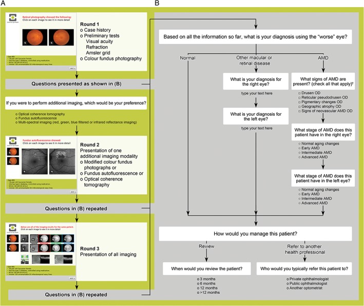
Figure 1.

A: Concept flowchart illustrating the questions and data captured in each case vignette (computer‐based case simulation). Each case is presented as a clinical scenario in a slide‐show format whereby the participant first reads about the patient's case history and preliminary test findings (visual acuity, refraction and Amsler grid). Colour fundus photographs are then presented and the participant queried regarding their diagnosis, management and imaging preferences (round one). B: The questions (other than imaging preference) are repeated following presentation of images using one imaging modality (round two), and then again following presentation of all imaging results (round three). Participants had the option of reviewing the fundus photographs at any time. †Response options for the left eye were also provided. AMD: age‐related macular degeneration, OD: right eye.
