
Figure 4.
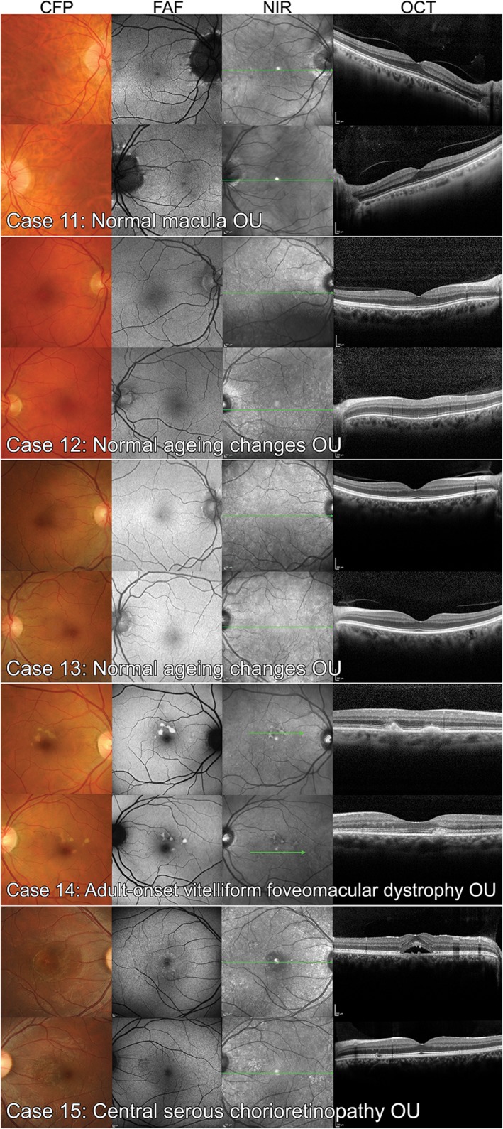
Non‐age‐related macular degeneration case images of the six normal eyes (four with normal ageing changes) and four eyes with other macular diseases used in the study. CFP: colour fundus photography, FAF: fundus autofluorescence, NIR: near infrared reflectance, OCT: optical coherence tomography.