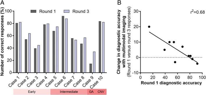
Figure 7.

A: Diagnostic accuracy as a function of the case type and round. Round one responses were based on colour fundus photography alone, while round three followed the review of colour fundus photographs and all of the advanced imaging findings. Across the cohort, 7/10 cases showed an improvement in diagnostic accuracy ranging from one per cent to 20 per cent. Three out of 10 cases (cases 6, 7 and 10) showed a decrease in diagnostic accuracy between minus one per cent and minus four per cent. The decrements were associated with cases that had high diagnostic accuracy using colour fundus photography alone. B: Scatterplot demonstrating the correlation between round one diagnostic accuracy using colour fundus photography and the improvement with multimodal imaging. CNV: choroidal neovascularisation, GA: geographical atrophy.
