
Figure 5.
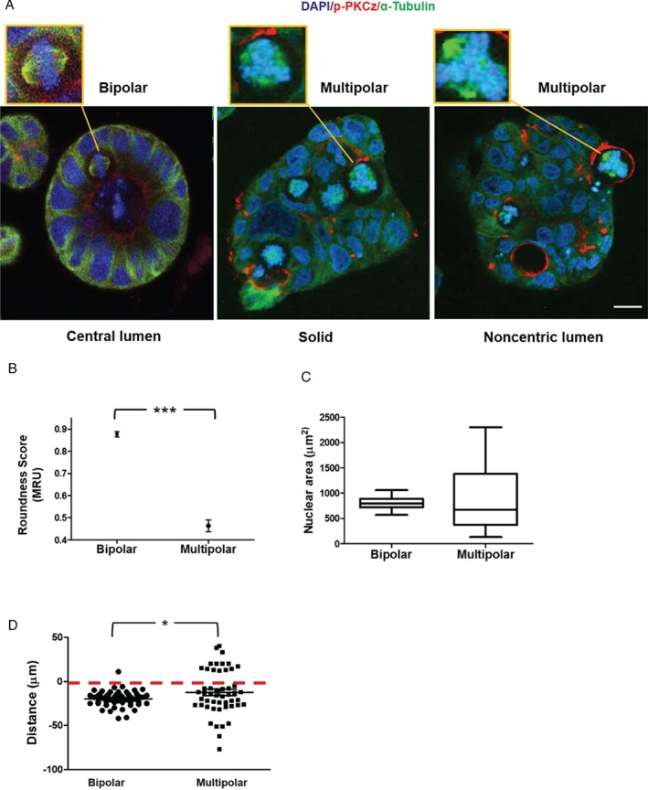
Relationships between mitotic spindle geometry and multicellular morphology in 3D organotypic CRC cultures. (A) Confocal assays of spindle architecture (insets) and multicellular morphology in 3D organotypic cultures. Control Caco‐2 cultures with appropriately orientated bipolar spindles (left panel) had regular 3D morphology with single central lumens surrounded by a uniform apical membrane and columnar epithelial monolayers. SiRNA knockdown of PKCz in 3D Caco‐2 PLK4OE cultures induced multipolar spindle formation and solid cell‐filled 3D structures with dispersed apical membrane foci. These cultures either lacked any lumen (middle panel) or had aberrant noncentric lumens lying outwith gland centres, surrounded by atypical epithelium (right panel). Cells with multipolar spindles extended across the basal interface with extracellular matrix (ECM) more frequently than cells with bipolar spindles (see D). (B) Nuclear ‘roundness’ scores in glands with bipolar versus multipolar spindles. ***p < 0.001; paired Student's t‐test. A score of 1 MRU denotes a perfect circle 70 (n = 60 cells from glands containing bipolar or multipolar spindles). (C) Range of nuclear size in glands with bipolar versus multipolar spindles (p < 0.01; Levene's test; n = 30 bipolar or multipolar cells). (D) Summary extension of cells with bipolar or multipolar spindles across the ECM interface (denoted by red interrupted line). Distances between spindle midpoints and the ECM interface were assessed. Positive or negative values were assigned for direction of extensions into or away from the ECM, respectively. Positive distance values (into the ECM) in multipolar versus bipolar spindles = 14/50 versus 1/50; *p = 0.03; paired Student's t‐test. Staining: DAPI blue, for nuclear DNA; p‐PKC red, for apical membranes, α‐tubulin green, for microtubules. Assays at 4 days of 3D culture.