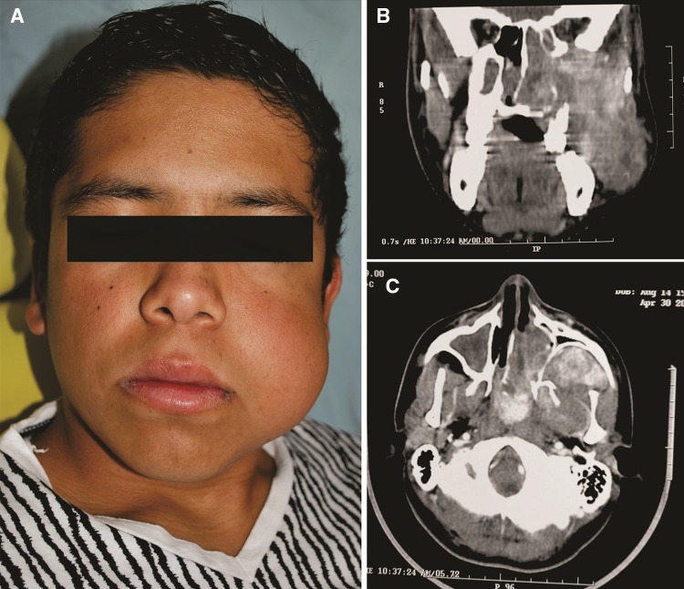
Fig. 1.
14-year-old male with a tumoral mass affecting the nasopharynx and extending into left cheek. a Clinically the patient shows facial left swelling and asymmetry. This clinical aspect was favored by a previously unsuccessful attempt of surgical removal. b In a coronal CT section, a large tumoral mass deviating the nasal septa and extending from sinonasal spaces to pterygopalatine fossa, infratemporal fossa and extents into soft tissues of the check. c Axial CT image of the same tumor shows anterior displacement of posterior wall of left maxillary sinus is evident. Contrast media highlights the prominent vascularity

