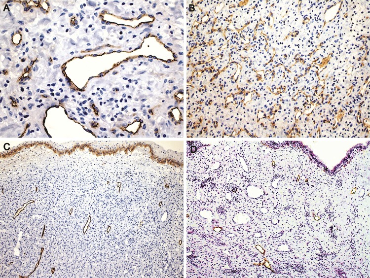
Fig. 5.
Immunohistochemical features. a Varying size vessels showing flat endothelial cells (CD34, ×400). b CD31 expression in area rich in microvessels (×200). c, d Lymphatic vessels localized exclusively in regions near the surface epithelium. The number of lymphatic vessels is variable. Note the squamous metaplasia of the epithelium with basal epithelial cells positive for podoplanin serving as internal control in figure c (Podoplanin, ×200)

