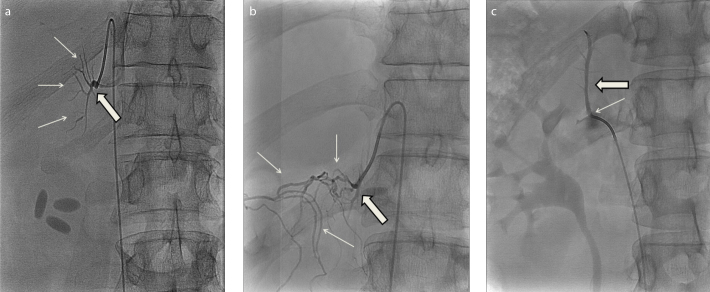
Figure. a–c.
Radiographic images from adrenal catheterizations showing normal anatomy of the right adrenal vein (a, thick arrow) draining with a sharp angle into the cava inferior approximately 2 cm above the orifice of the left renal vein, as well as small branches from the adrenal vein (a, thin arrows); anatomical variation of the right adrenal vein (b, thick arrow) with several collateral veins of unknown origin (b, thin arrows); and a rare anatomical variation in which the right adrenal vein (c, thick arrow) drains into the right renal vein near its orifice into the vena cava (c, thin arrow).

