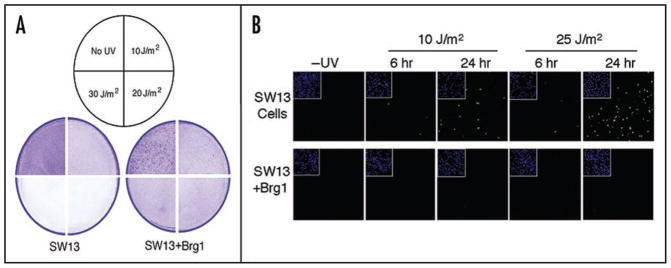
Figure 3.
Sensitivity of SW13 and SW13 + Brg1 cells to UV radiation. (A) Cell detachment assay. Monolayers of cells (~80% confluent) were exposed to various doses of UV radiation (shown in sectored circle), incubated for 24 hr and fixed/stained with ethanol/crystal violet. Figures depict quadrants from plates exposed to the different UV doses. (B) Brg1 expression in SW13 cells suppresses UV induced apoptosis. SW13 and SW13 + Brg1 cells were UV irradiated (10 or 25 J/m2), incubated for 6 hr or 24 hr as indicated, and analyzed using the TUNEL assay and confocal microscopy. Insets show DAPI staining of nuclei.

