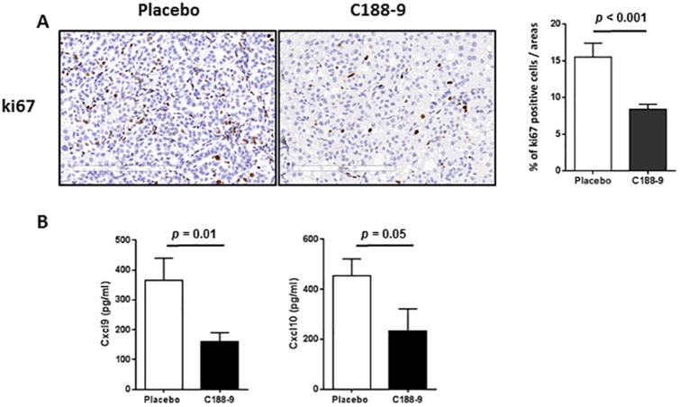
Figure 6. Effects of C188-9 treatment on tumor cell proliferation and chemokine levels in serum.
In both vehicle and C188-9-treated mice, ki67 immunohistochemical (IHC) staining was used to evaluate the amount of tumor cell proliferation. (A) Representative sections are shown. Scale Bars, 200 μm. (B) Quantitation of IHC-positive staining for ki67 expressed as percentage of positive tumor cells. (C) Serum levels of CXCL9 and CXCL10 in vehicle- and C188-9-treated HepPten-mice. Values represent mean ± SEM (Mann-Whitney test).

