Figure 3.
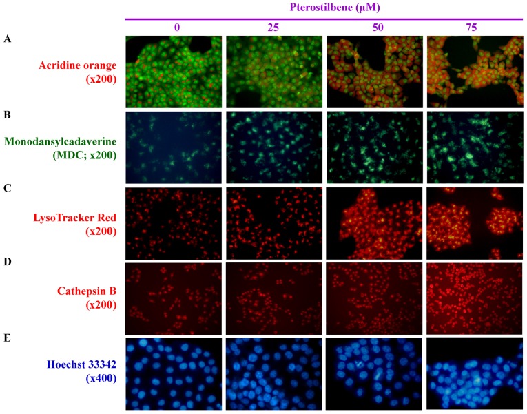
Effects of pterostilbene on the autophagy and DNA condensation of CAR cells. The cells were treated with 0, 25, 50 and 75 μM pterostilbene for 24 h and then probed using (A) acridine orange to detect acidic vesicular organelles, indicated by a red color (magnification, ×200). (B) Monodansylcadaverin, an autophagolysosome marker, indicated by a green color (magnification, ×200). (C) LysoTracker Red to determine lysosomal function, indicated by a red color (magnification, ×200). (D) Cathepsin B to detect lysosomal activity, indicated by a red color (magnification, ×200). (E) Hoechst 33342 staining to observe cell nuclei, as indicated by a blue color (magnification, ×400). Representative images were taken from three independent experiments.

